To gain access to the lower portions of the canal It also facilitates placement of topical medications in the external ear canal.įor patients with otitis externa where there is minimal hyperplasia of the epithelium of the external auditory meatusįor removal of small mass lesions of the lateral aspect of the vertical canal Lateral wall resection (LWR) increases drainage and improves ventilation of the ear canal.

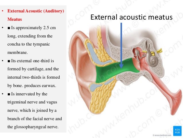
The terminal portion of the horizontal canal is formed by a bony projection of the petrous temporal bone.Ĭlick on the figure to see a larger view. It comprises a vertical canal and a horizontal canal. The external ear canal is a funnel-shaped cartilaginous tube which extends from the external auditory meatus to the tympanic membrane. Books & VINcyclopedia of Diseases (Formerly Associate).VINcyclopedia of Diseases (Formerly Associate).A confident diagnosis in such scenarios is aided by the presence of accompanying bone destruction and absent contrast enhancement. To conclude, external auditory canal cholesteatoma can have atypical imaging appearances as hyperdense lesions with high HU values. Mycetoma of the mastoid with bony erosions are described, but extensive disease with ossicular erosions are not reported. The features which made keratosis obturans less likely was the chronic history and the irregular bony erosions instead of the smooth scalloping seen in keratosis. Keratosis obturans is the pathological condition characterised by accumulation of desquamated keratin in the external auditory meatus. The imaging differentials include keratosis obturans and mycetoma.

The features of facial palsy alerted the ENT surgeon to a pathology not confined to the external auditory canal but extending to the middle ear. Our patient had a previous history of trauma which could be the inciting event for the secondary cholesteatoma formation. Īn external auditory canal cholesteatoma can be mimicked by many conditions like keratosis obturans, carcinoma etc, computed tomographic imaging of the temporal bone is useful in arriving at the correct diagnosis, to determine the extent of the lesion and the bony involvement. In case of involvement of the mastoid air cells as in our case, the appropriate procedure should be canaloplasty with mastoidectomy. The management of the external auditory canal cholesteatoma depends on the extent of the lesion. The case under discussion is in stage III. There is extension beyond the temporal bone in stage IV. In stage III there is involvement of mastoid air cells. There is involvement of tympanic membrane and middle ear in stage II. In stage I, the disease limited to the external auditory canal.

However, a homogenously hyperdense appearance is not described which is peculiar to our case.Īccording to the clinical and computed tomography staging by Seung-Ho Shin et al there are four groups of external auditory canal cholesteatoma.

Ĭholesteatoma of the external auditory canal appears as a soft tissue attenuating lesion with bone erosion and with intramural bone fragments. Usual symptoms include otalgia, otorrhea and hearing loss. Most of the cases are spontaneous but can occur secondary to trauma or surgery. External auditory canal cholesteatomas are rare occurring in about 1-5 of every 1000 otologic cases. Cholesteatomas occur due to the ingrowth of the stratified squamous epithelium of the external auditory canal into the middle ear.


 0 kommentar(er)
